Michael Giakoumis: Physiotherapist: British Athletics, Total Performance & Centre of Health and Human Performance (London)
Carles Pedret MD, PhD: Sports Medicine and Imaging department. Clinica Diagonal (barcelona)
Sprinting places a significant demand on the rectus femoris (RF) and is one of the most common mechanisms of injuries for this muscle (Hasselman et al., 1995). During the initial swing phase in sprinting the hip moves into flexion reaching a peak angular velocity of 792ºs-1. The shank, which is moving into flexion, reaches a peak angular velocity of 1185ºs-1 when sprinting at 9.9ms-1 (Miyashiro et al., 2019). The RF contributes to hip flexion torque following toe off and works to decelerate the knee flexion angular velocity around the same period of time. Research shows that the intramuscular forces that the RF produces exceeds 2000N in an 80kg individual running at 9ms-1(Dorn et al., 2012). Therefore in the context of this case study, a sub 10s sprinter capable of reaching a top speed of 11.8ms-1, the intramuscular forces can be anticipated to be significantly larger. These intramuscular forces also occur during peak muscle activation and length. This places a significant strain on the passive structures such as the intramuscular and free tendon therefore increasing the risk of injury on the non-contractile tissue.
Proximal Anatomy of the Rectus Femoris
The proximal anatomy of the RF consists of two-to-three free tendons; the direct head, originating from the anterior inferior iliac spine, an indirect or reflected head, originating from the anterior capsule, lateral margin of the superior acetabular ridge and lateral acetabular rim of the hip joint, and in the majority of individuals a third head that has two primary attachments, one to the iliofemoral ligament and the other the gluteus minimus muscle (Ouellette et al., 2006; Tubbs et al., 2006). The anatomy alone suggests the RF has a direct and indirect role in providing active and passive stability to the hip but also may be prone to shearing injuries at the conjoining intersection (reference). The direct and indirect tendons conjoin but act separately just below the AIIS (Kassarjian et al., 2012) forming two distinctively different structured muscles but are cadaverically indistinguishable. The image of a hotdog bun with a sausage in between may help to visualise the structure of the RF. The outer-layered muscle (or bun) has fibres running from the anterior to posterior aponeurosis forming a unipennate muscle and is associated with the direct head tendon. These muscle fibers wrap around the muscle fibers in the middle that are originating from the indirect heads central tendon. Muscle fibers originate from either side of the indirect heads central tendon and insert on the fascia and posterior aponeurosis forming a bipennate muscle.
The role of a free tendon is to transfer force from the muscle across a joint. Additionally, most free tendons of the lower limb act as springs and the RF is no different (Lai et al., 2019). Its architecture suggests it is most effective in transferring force during high-speed running as its long tendon slack length is taken up optimising its function. Therefore in order to ensure its tendon can express its tendon like behaviour, an appropriate level of force and active muscle stiffness is required. Thus in any injury, having knowledge of the anatomy can help understand the injury and influence the rehabilitation process.

Figure 1. Rectus Femoris (Cadaveric Representation)

Figure 2. Rectus Femoris
Case Study
Below is a case study of a 100m international sprinter who sustained an acute injury to his RF. On initial MRI findings it was demonstrated that the injury was consisted of a high-energy injury. There was a partial transverse tear through both his indirect and direct proximal tendons with an associated split between the two tendons at their conjoining junction (Figures 3.). Using the British Athletics Muscle Injury Classification (BAMIC) system (Pollock et al., 2014), this was diagnosed as a 3C injury to the RF muscle involving the free tendon and intramuscular tendon.
Progression through rehabilitation should be based on healing time frames entwined with clinical milestones for each phase of the rehab process. However, there is a lack of evidence describing criteria for progression in RF injuries. Using experience, extrapolating information from biomechanical literature on the demands on the tissue and combining this with the demands of the task can help provide the necessary milestones for return to sprinting and performance.
The most important phase(s) are the progression towards transitioning from running to accelerating/sprinting. During these periods an increase in force and strain on the RF occurs to which the series elastic components ability to tolerate and transfer forces, with high tensile loads on the tendon and muscle connective tissue, is vital. As running speed increases this strain behavior increases, as does the amount of negative work. Thus, ensuring all milestones are aligned to building tolerance and appropriate adaptation for this phase from the onset is a key aspect of RF rehabilitation.
As with the above process, undergoing a gap analysis in order to understand the individual’s risk factors that lead to an increased risk of injury should be addressed. Those that pertain to adaptable physical qualities can then be targeted under an adaptation based model rather than a clinical recipe of progression.
Below a 7-stage rehabilitation framework is demonstrated that progressed and prepared the individual for the demands of the activity moving from a muscle specific to functional specific progression.
Principles of Rehabilitation

Stage 1. Early Management
The initial 48 hours are crucial in the healing and remodeling process. The athlete was encouraged to protect the muscle and keep it in a shortened position. Compression and ice using the Game Ready was implemented to reduce pain and swelling.
Stage 2. Early Loading & Contributing Factors
Intramuscular tendons and free tendons may take up to 6 weeks or longer for complete histological healing (Jaspers et al., 2005). Therefore loading in positions with less tensile stress such as hip flexion with knee extension then progressing to mid-range (anatomical neutral) was used in order to respect the healing tissue. The principle of lateral force transmission in the early loading phase was also utilised (Bloch and Gonzalez-Serratos, 2003). This principle safely loads the tissue as force from other contracting muscles is inter-muscularly transmitted to the target tissue. This reduces the tensile stress on the tissue but allows force to be applied across the target tissue in a safe position whilst still leading to mechanotransduction. Hip adduction and abduction exercises whilst in knee extension in varying hip flexion positions are examples and were used within the first 10-14 days.
Stress relaxation a phenomenon that increases the stress on injured tissue is a mechanism to be mindful of (Baar, 2019). If load is constantly held for long periods of time (30s+), the tissue undergoes creep and diverts stress towards the injured tissue. Because the scar is still immature and weak the excessive stress on the healing tissue may not be advantageous and therefore shorter duration isometrics were utilised.
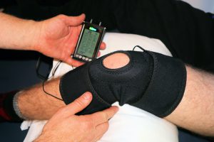
Blood Flow Restriction (BFR) with electrical stimulation (E-Stim) was used as an adjunct form of loading in the initial stages. It provides an alternative loading strategy that can help limit the negative neuromuscular effects following injury. BFR+E-Stim aims to limit muscular atrophy, provide an aerobic stimulus, and stimulate slow and fast twitch fibres to maintain activity. With the combination of the two modalities, local muscle fatigue can occur, therefore through Henneman’s size principle, a progression from slow to fast twitch muscle fibre utilization will occur if the stimulus is provided long enough towards volutional fatigue. Because of the low activation and suggested long duration, the stimulus will require aerobic metabolism to provide the energy for muscle contraction (Pearson and Hussain, 2015). This will help improve the oxidative capacity of the muscle where increases in muscle capillarization, increased concentration of oxidative enzymes, and an increase in mitochondrial density will occur (Laursen & Bucheitt 2018).
Following a thorough assessment within the constraints of the injury, other potential contributing factors were identified and subsequently addressed in stages 2 and 3. These were thought to act as internal risk factors and included a reduction in calf function (Farris et al., 2019; Lewis and Ferris, 2008), an increase in rotational movement at the knee (Flaxman et al., 2012) and a reduction in hip flexion and adduction strength relative to hip extension strength (Neumann, 2010).
Stage 3: Symmetrical Loading, Strength Endurance & Peak Force
The quadriceps as a muscle group are key in both absorbing and generating vertical force and power during stance phase (Dorn et al., 2012). Having vertical force capability will help in the ability to run up to 7m/s (Dorn et al., 2012). In order to optimize vertical forces, high loads are required which cannot be achieved to the same extent during unilateral tasks. These high loads utilized will help to develop strength throughout the kinetic chain (compound movements) from the trunk, to the hips, to the foot and ankle complex and through the force transferring nature of the bi-articular muscles. Alongside improving the vertical force capability stage 3 also aims to restore strength and capacity of the injured tissue.
In this case study individual examples were high volume based training of narrow ¼ squats and eventually small box step ups when large forces on the tissue were warranted. The progression of building up capacity first whilst the tissue was still healing (5×15 @ 160kg) then moving towards stress testing the tissue with high intensity exercises (5 sets of 3 @ 180kg step ups) was the progression. These exercises when coached appropriately bias the knee to be the primary contributor to vertical movement.
Stage 4: Unilateral Loading & Strain
Stage 4 covers the development for the capacity to produce speed and aims to improve unilateral torque and the reciprocal movement of flexion and extension that is required to increase stride frequency. Unilateral loading progresses the ability to develop unilateral strength and optimize tissue development whilst also allowing larger ranges of movement. Site-specific adaptations can be induced by training either the distal or proximal segment. If distal adaptations are required, loading at the knee induces greater adaptations distally, whereas the opposite is true for the proximal segment especially in regards to RF (Ema et al., 2018; Noorkõiv et al., 2015). This information was applied by using the total hip machine to induce stress towards the hip flexors, RF included.
In this phase progressing the degree of strain and strain rate may be an important variable to consider in order to restore the viscoelastic properties of the musculotendinous unit. For strain and strain rate, isometric and eccentric contractions at longer lengths were utilized to improve the viscoelastic properties by upregulating adhesion molecules, collagen synthesis and influencing the material properties’ (Arampatzis et al., 2007; Franchi et al., 2017). Because of the tendon split, eccentric front leg elevated split lunges were used as these have been shown to induce higher levels of muscle force in the RF compared to other exercises whilst achieving the appropriate molecular response in the local tissue. As the athlete was able to lift 105kg, the RF intramuscular force was calculated to reach approximately 4810N (Schellenberg et al., 2017). Sustained isometrics may also be beneficial to utilize the stress-relaxation principle and creep phenomenon, inducing greater degrees of strain on the non-contractile tissue (Baar, 2019). This is the stage where this phenomenon is best utilized. As the free tendons were shown to have continuity (subsequent MRI) but yet still remodeling, stressing that tissue would enhance the remodeling process and restore the viscoelastic properties.
Stage 5: Return to Run & Rate of Loading
Stage 5 involves gym-based loading that progresses the individual along the force-velocity spectrum incorporating power (force x velocity) and speed-based movements. These activities will help to increase the neural drive and coordination of the neuromuscular system.
Although development of the non-contractile tissue is likely to have occurred during stages 3 and 4 with the use of isometrics and eccentrics at longer lengths, further development and preparation for short-stretch cycles will be important. Therefore, conditioning of this tissue should be centred on fatiguing concentric based actions in order to increase the reliance and hence adaptations on the non-contractile tissue when SSC exercises are performed. An example is video X.
Concurrently or preceding returning to on feet running, power based activity in the gym should be commenced. Activities such as weighted and un-weighted counter-movement jumps are a way to facilitate RFD adaptations that will enhance the phasic muscle activation patterns required. Furthermore this may induce architectural adaptations to both the proximal and distal segments of RF as well as facilitate adaptations specific to its fibre type distribution (Blazevich et al., 2003; Johnson et al., 1973).
Progressing to hill runs, ‘Mach’ drills such as A skip and B skip and backwards running can help condition the quadriceps gradually exposing them to increasing levels of strain prior to high-velocity sprinting. These exercises will improve the inter-muscular coordination, proximal-distal coordinative sequencing, firing frequency and agonist co-activation that are required for optimal bi-articular muscle function.
For this case study, the individual was progressed from walking drills, to dynamic drills, to backwards running, then dribbles, and finally strides as a progression prior to return to sprinting. For plyometric progressions we used pogo circuits progressing to CMJ’s, rapid hip flexion on a decline board then to alternating lunge jump switches and Split Lunge Jump Scissors.
Stage 6: Return to Sprinting
There is nothing that can replicate the task of sprinting and the neuromuscular demands and speed of movement other than sprinting itself. Stage 6 is an extension of stage 5 and the proceeding stages before. By this stage the individual had first regained the required capacity, force, power, and then finally speed of movement prior to increasing running velocity. Within this stage the kinematics and technical model of the coach was of a large emphasis. Biomechanical analysis assessing step length and ground contact time alongside kinematic analysis was utilized to support the assessment of symmetry and peak hip flexion and extension angles and any underlying neuromuscular guarding at speed. These data points were then referenced against pre-injury data.
Throughout the stages the coach of the athlete was always consulted on and involved with the process however their responsibility and influence over the program continued to grow. It is at this stage where the coach was making the majority of decisions and only supplementary work was provided by the medical team.
Stage 7: Return to Performance
As it has been stressed especially in stages 3 and 4, this framework is centred on improving the individual’s health and human performance which covers addressing the local pathology and contributing factors. Stage 7 is a confirmation stage to which the athlete should have completed all tasks required to perform. Prior stages would have laid the appropriate foundations of not only the local tissue injured, but all required attributes of the athlete.
Progressing through stages in our opinion requires both time based and objective and functional based markers. Knowledge of the pathophysiological processes during healing and the minimum and ideal benchmark requirements for both the local tissue and kinetic chain is important and must be made relevant to the individual and the demands of the sport/task. It is therefore recommended to start with the end goal in mind and work backwards to first of all appreciate the speed of movement, the force demands but also the repeated demands of each step in a 100m sprint. Ultimately the performances of an individual are multidimensional but in a linear and pure sport of athletics performance can be easily measured. At the time of this article due to the recent nature of the injury competitive performance results cannot be provided so this is to be confirmed.
Milestones:

Acknowledgements:
The current and futures success of this case study could not be achieved without the support from Dr James Brown, Dr Noel Pollock and Helen Van Kempen from British Athletics as well as Gordon Bosworth and Coach Ryan Freckleton.
We would also like to acknowledge the below for providing the cadaveric image.
Dr. Francisco Reina de la Torre. MD. PhD.
Dra. Anna Carrera Burgaya.MD. PhD.
NEOMA Research Group.
Medical Science Department. Faculty of Medicine. Universitat de Girona.
Lastly we would like to thank the athlete for their dedication, diligence and focus during the rehabilitation process as without this the current outcome would not have been achieved.
Arampatzis, A., Karamanidis, K., Albracht, K., 2007. Adaptational responses of the human Achilles tendon by modulation of the applied cyclic strain magnitude. J. Exp. Biol. 210, 2743–2753. https://doi.org/10.1242/jeb.003814
Baar, K., 2019. Stress Relaxation and Targeted Nutrition to Treat Patellar Tendinopathy. Int J Sport Nutr Exerc Metab 29, 453–457. https://doi.org/10.1123/ijsnem.2018-0231
Blazevich, A.J., Gill, N.D., Bronks, R., Newton, R.U., 2003. Training-specific muscle architecture adaptation after 5-wk training in athletes. Med Sci Sports Exerc 35, 2013–2022. https://doi.org/10.1249/01.MSS.0000099092.83611.20
Bloch, R.J., Gonzalez-Serratos, H., 2003. Lateral force transmission across costameres in skeletal muscle. Exerc Sport Sci Rev 31, 73–78. https://doi.org/10.1097/00003677-200304000-00004
Dorn, T.W., Schache, A.G., Pandy, M.G., 2012. Muscular strategy shift in human running: dependence of running speed on hip and ankle muscle performance. J. Exp. Biol. 215, 1944–1956. https://doi.org/10.1242/jeb.064527
Ema, R., Saito, I., Akagi, R., 2018. Neuromuscular adaptations induced by adjacent joint training. Scand J Med Sci Sports 28, 947–960. https://doi.org/10.1111/sms.13008
Farris, D.J., Kelly, L.A., Cresswell, A.G., Lichtwark, G.A., 2019. The functional importance of human foot muscles for bipedal locomotion. Proc Natl Acad Sci U S A 116, 1645–1650. https://doi.org/10.1073/pnas.1812820116
Flaxman, T.E., Speirs, A.D., Benoit, D.L., 2012. Joint stabilisers or moment actuators: the role of knee joint muscles while weight-bearing. J Biomech 45, 2570–2576. https://doi.org/10.1016/j.jbiomech.2012.07.026
Franchi, M.V., Reeves, N.D., Narici, M.V., 2017. Skeletal Muscle Remodeling in Response to Eccentric vs. Concentric Loading: Morphological, Molecular, and Metabolic Adaptations. Front Physiol 8. https://doi.org/10.3389/fphys.2017.00447
Hasselman, C.T., Best, T.M., Hughes, C., Martinez, S., Garrett, W.E., 1995. An explanation for various rectus femoris strain injuries using previously undescribed muscle architecture. Am J Sports Med 23, 493–499. https://doi.org/10.1177/036354659502300421
Jaspers, R.T., Brunner, R., Riede, U.N., Huijing, P.A., 2005. Healing of the aponeurosis during recovery from aponeurotomy: morphological and histological adaptation and related changes in mechanical properties. J. Orthop. Res. 23, 266–273. https://doi.org/10.1016/j.orthres.2004.08.022
Johnson, M.A., Polgar, J., Weightman, D., Appleton, D., 1973. Data on the distribution of fibre types in thirty-six human muscles. An autopsy study. J. Neurol. Sci. 18, 111–129. https://doi.org/10.1016/0022-510x(73)90023-3
Kassarjian, A., Rodrigo, R.M., Santisteban, J.M., 2012. Current concepts in MRI of rectus femoris musculotendinous (myotendinous) and myofascial injuries in elite athletes. Eur J Radiol 81, 3763–3771. https://doi.org/10.1016/j.ejrad.2011.04.002
Lai, A.K.M., Biewener, A.A., Wakeling, J.M., 2019. Muscle-specific indices to characterise the functional behaviour of human lower-limb muscles during locomotion. J Biomech 89, 134–138. https://doi.org/10.1016/j.jbiomech.2019.04.027
Lewis, C.L., Ferris, D.P., 2008. Walking with Increased Ankle Pushoff Decreases Hip Muscle Moments. J Biomech 41, 2082–2089. https://doi.org/10.1016/j.jbiomech.2008.05.013
Miyashiro, K., Nagahara, R., Yamamoto, K., Nishijima, T., 2019. Kinematics of Maximal Speed Sprinting With Different Running Speed, Leg Length, and Step Characteristics. Frontiers in Sports and Active Living 1, 37. https://doi.org/10.3389/fspor.2019.00037
Neumann, D.A., 2010. Kinesiology of the hip: a focus on muscular actions. J Orthop Sports Phys Ther 40, 82–94. https://doi.org/10.2519/jospt.2010.3025
Noorkõiv, M., Nosaka, K., Blazevich, A.J., 2015. Effects of isometric quadriceps strength training at different muscle lengths on dynamic torque production. J Sports Sci 33, 1952–1961. https://doi.org/10.1080/02640414.2015.1020843
Ouellette, H., Thomas, B.J., Nelson, E., Torriani, M., 2006. MR imaging of rectus femoris origin injuries. Skeletal Radiol 35, 665–672. https://doi.org/10.1007/s00256-006-0162-9
Pearson, S.J., Hussain, S.R., 2015. A review on the mechanisms of blood-flow restriction resistance training-induced muscle hypertrophy. Sports Med 45, 187–200. https://doi.org/10.1007/s40279-014-0264-9
Pollock, N., James, S.L.J., Lee, J.C., Chakraverty, R., 2014. British athletics muscle injury classification: a new grading system. Br J Sports Med 48, 1347–1351. https://doi.org/10.1136/bjsports-2013-093302
Schellenberg, F., Taylor, W.R., Lorenzetti, S., 2017. Towards evidence based strength training: a comparison of muscle forces during deadlifts, goodmornings and split squats. BMC Sports Sci Med Rehabil 9, 13. https://doi.org/10.1186/s13102-017-0077-x
Tubbs, R.S., Stetler, W., Savage, A.J., Shoja, M.M., Shakeri, A.B., Loukas, M., Salter, E.G., Oakes, W.J., 2006. Does a third head of the rectus femoris muscle exist? Folia Morphol (Warsz) 65, 377–380.


